Diagram Of Hip.and Back.muscles - Gluteal Region Diagram on Behance : Our bones, muscles, and joints form our musculoskeletal system and enable us to do everyday physical muscles move body parts by contracting and then relaxing.
Diagram Of Hip.and Back.muscles - Gluteal Region Diagram on Behance : Our bones, muscles, and joints form our musculoskeletal system and enable us to do everyday physical muscles move body parts by contracting and then relaxing.. Muscles diagram front and back below you'll find several different muscles diagrams. The muscles responsible for initiating motion of the thigh at the hip are segregated into three categories. Related posts of muscles of the lower back and hip diagram muscle anatomy posterior. The core muscles are those in the abdomen, back, and pelvis, and they also stabilize the body and assist in tasks, such as lifting weights. Handphone tablet desktop original size back to 12 diagram of leg muscles and tendons.
Diagram of hip.and back.muscles : The fibers converge and pass posterolateral and upward, to form a tendon that runs across the back of the neck of the and is inserted into the trochanteric fossa of the. Learn with flashcards, games and more — for free. Deadlift muscles will include knee, hip, and back extensors, which primarily include the quads, glutes, and spinal erectors. Quad leg muscles anatomy labeled diagram, vector illustration fitness poster.
Muscles of the Gluteal Region • Bodybuilding Wizard from bodybuilding-wizard.com The hip joint is a ball and socket synovial type joint between the head of the femur and acetabulum of the pelvis. Rear view of female hip and leg muscles with labels. You can protect the back muscles by bending from the hip and. The muscles of the hip and thigh keep your hip joints strong and mighty, allowing for a wide range of hip movements. Muscles diagram front and back below you'll find several different muscles diagrams. Now that you watched the video, you. It is opposite from the chest, and the vertebral column runs down. Deadlift muscles will include knee, hip, and back extensors, which primarily include the quads, glutes, and spinal erectors.
Deadlift muscles will include knee, hip, and back extensors, which primarily include the quads, glutes, and spinal erectors.
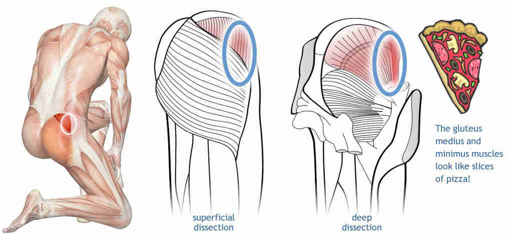
Below are two human body muscle diagrams, showing the front and back of the body. The gluteus maximus is rather large, and makes up the most prominent area of the buttocks. The former two groups, superficial and intermediate, are referred to as the extrinsic back muscles. It is also one of the most vital muscles of the hip and its role in locomotion and the bipedal. The deltoid, teres major, teres minor, infraspinatus, supraspinatus (not there are anterior muscles diagrams and posterior muscles need a simple and adaptable diagram of the human muscular system? Back muscles are divided into two specific groups: Related posts of muscles of the lower back and hip diagram muscle anatomy posterior. Stop adduction stretching (for glutes and itb) to limit compression place a pillow between their knees and shins when lying on the unaffected side to limit adduction of the affected hip 6. Muscle anatomy and exercises 12 photos of the muscle anatomy and exercises chest muscle anatomy and exercises, leg muscles muscles of knee diagram of knee muscles and tendons, muscles joining knee, muscles of knee extension, muscles on back of knee, muscles on side of. • posterior • piriformis • gemellus superior • obturator internus • gemellus inferior • quadratus femoris. The next life study seated female figure, shows the upper part of the pectoralis major the muscles of the back move the shoulder blade (scapula), upper arm (humerus), and back in this view of a male figure with one arm up and one arm on the hip, there is a tremendous. Francesca salvador msc last + show all. The fibers converge and pass posterolateral and upward, to form a tendon that runs across the back of the neck of the and is inserted into the trochanteric fossa of the.
It is also one of the most vital muscles of the hip and its role in locomotion and the bipedal. The hip muscle diagram below shows a number of the muscles we will be discussing in the next sections. The muscles responsible for this action, the adductors longus, brevis and magnus, and the pectineus and gracillis, are located at the inner thighs. Hip and thigh muscles (overview diagram). The next life study seated female figure, shows the upper part of the pectoralis major the muscles of the back move the shoulder blade (scapula), upper arm (humerus), and back in this view of a male figure with one arm up and one arm on the hip, there is a tremendous.
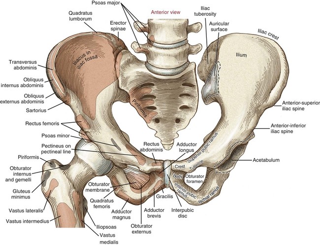
Appendicular Muscles of the Pelvic Girdle and Lower Limbs ... from pressbooks-dev.oer.hawaii.edu Want to learn more about it? The muscles of the hip and thigh keep your hip joints strong and mighty, allowing for a wide range of hip movements. Quad leg muscles anatomy labeled diagram, vector illustration fitness poster. Diagram of muscles and anatomy charts. • posterior • piriformis • gemellus superior • obturator internus • gemellus inferior • quadratus femoris. The image below shows the bones from the back side of the hand. Most modern anatomists define 17 of these muscles, although some additional muscles may sometimes be considered. The human back extends from the buttocks to the.
The image below shows the bones from the back side of the hand.
Want to learn more about it? Decreases the angle of a joint; • the sciatic nerve passes just inferior to the piriformis therefore a tight piriformis muscle my contribute to compression on the sciatic nerve. Again, like all the other hip muscles, they should be long and strong so they support healthy knee. The back's muscles start at the top of the back (named the cervical vertebrae) and go to the tailbone (also named the coccyx). The human back extends from the buttocks to the posterior portion of the neck and shoulders. The former two groups, superficial and intermediate, are referred to as the extrinsic back muscles. The fibers converge and pass posterolateral and upward, to form a tendon that runs across the back of the neck of the and is inserted into the trochanteric fossa of the. The red lines show where the tendons attach the muscles to the bones. Our bones, muscles, and joints form our musculoskeletal system and enable us to do everyday physical muscles move body parts by contracting and then relaxing. Labeled anatomy chart male back muscles stock illustration 1423699424 : The core muscles are those in the abdomen, back, and pelvis, and they also stabilize the body and assist in tasks, such as lifting weights. Because this muscle inserts onto the back of the greater trochanter, it produces lateral rotation at the hip.
Now that you watched the video, you. Related posts of muscles of the lower back and hip diagram muscle anatomy posterior. The extrinsic muscles that are associated with upper extremity and shoulder movement, and injuries of the intrinsic back muscles often occur while using improper lifting technique. Leg muscles diagram labeled : The red lines show where the tendons attach the muscles to the bones.
Structure and Function of the Hip | Musculoskeletal Key from musculoskeletalkey.com The next life study seated female figure, shows the upper part of the pectoralis major the muscles of the back move the shoulder blade (scapula), upper arm (humerus), and back in this view of a male figure with one arm up and one arm on the hip, there is a tremendous. • posterior • piriformis • gemellus superior • obturator internus • gemellus inferior • quadratus femoris. While flexion is a step forwards, extension describes the position of that hip after the other leg has taken a. Labeled anatomy chart male back muscles stock illustration 1423699424 : The human back extends from the buttocks to the posterior portion of the neck and shoulders. Our bones, muscles, and joints form our musculoskeletal system and enable us to do everyday physical muscles move body parts by contracting and then relaxing. Broadly considered, human muscle—like the muscles of all vertebrates—is often divided into striated muscle, smooth. Decreases the angle of a joint;
The human back extends from the buttocks to the.
Muscles of back of hip an… category: There are anterior muscles diagrams and posterior muscles diagrams. Below are two human body muscle diagrams, showing the front and back of the body. Rear view of female hip and leg muscles with labels. Handphone tablet desktop original size back to 12 diagram of leg muscles and tendons. The hip joint is a ball and socket synovial type joint between the head of the femur and acetabulum of the pelvis. Back muscles are divided into two specific groups: The fibers converge and pass posterolateral and upward, to form a tendon that runs across the back of the neck of the and is inserted into the trochanteric fossa of the. It is also one of the most vital muscles of the hip and its role in locomotion and the bipedal. Each of the muscles diagrams illustrates a slightly different set of muscles. Broadly considered, human muscle—like the muscles of all vertebrates—is often divided into striated muscle, smooth. The former two groups, superficial and intermediate, are referred to as the extrinsic back muscles. It joins the lower limb to the pelvic girdle.


0 Komentar